

ePoster Viewing : Taewoo Kim
Airway Vista 2021
Longitudinal Alterations in Cement Dust Exposed Subjects via Quantitative Computed Tomography
Taewoo Kim1*, Myoung-nam Lim2*, Chang Hyun Lee3, Kum Ju Chae4, So Hyeon Bak5, Sung Ok Kwon5, Gong Yong Jin4, Eun-Kee Park6, Woo Jin Kim5†, and Sanghun Choi1†
1: Dept. of Mechanical Eng., Kyungpook University, 2: Biomedical Research Institute, Kangwon National University, 3: Dept. of Radiology, Seoul National University College of Medicine, Seoul National University Hospital, 4: Dept. of Radiology, Research Institute of Clinical Medicine of Jeonbuk National University Hospital, 5: Dept. of Internal Medicine and Environmental Health Center, Kangwon National University Hospital, 6: Dept. of Medical Humanities and Social Medicine, College of Medicine, Kosin University
1: Dept. of Mechanical Eng., Kyungpook University, 2: Biomedical Research Institute, Kangwon National University, 3: Dept. of Radiology, Seoul National University College of Medicine, Seoul National University Hospital, 4: Dept. of Radiology, Research Institute of Clinical Medicine of Jeonbuk National University Hospital, 5: Dept. of Internal Medicine and Environmental Health Center, Kangwon National University Hospital, 6: Dept. of Medical Humanities and Social Medicine, College of Medicine, Kosin University
Introduction: Cement dust exposure (CDE) has been known as one of potential risk factors, leading pulmonary diseases associated with an alteration of segmental airways and parenchymal lungs. We previously demonstrated that subjects with CDE had wall thickening, airway narrowing, and alteration of bifurcation angle, although their lung functions remained normal. With the longitudinal images for the same subjects, we aim to investigate temporal changes of quantitative computed tomography (QCT)-based imaging variables.
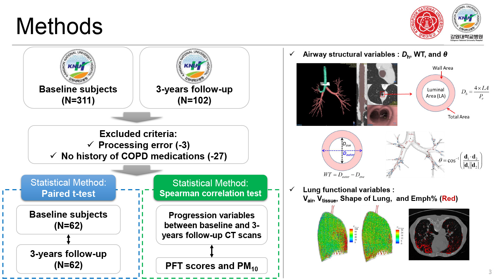
Methods: We obtained 302 CT scans of baseline subjects, and 102 CT scans of follow-up CDE subjects. Among the follow-up 102 data, three cases were failed during the image processing. With 62 CDE subjects with both baseline and follow-up QCT data excluding history of chronic obstructive pulmonary disease (COPD) medications, we respectively extracted QCT-based airway structures and lung functions. In airway structural variables, we extracted hydraulic diameter (Dh), wall thickness (WT), and bifurcation angle (θ). We also extracted air volumes (Vair), and tissue volumes (Vtissue) at total lung capacity (TLC), and functional residual capacity (FRC). Besides, we obtained Emphysema percent (Emph%), and functional small airway disease percent (fSAD%) with an image registration.
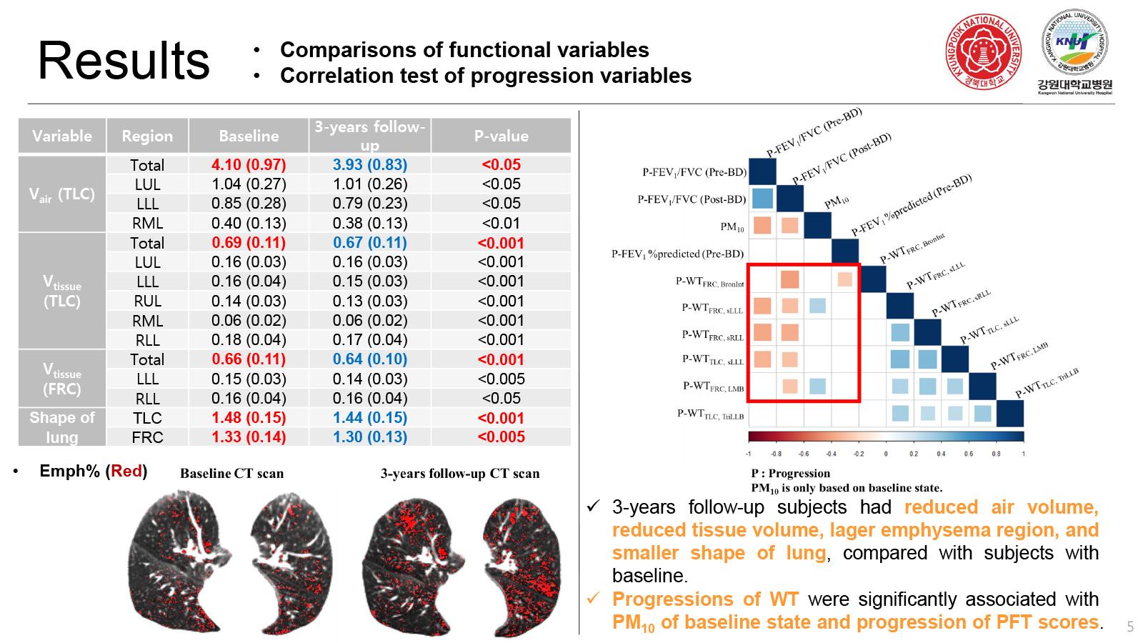
Results: Pulmonary function tests (PFT)’ and COPD assessment test values were within similar or even better in the follow-up cases, compared with baseline cases. Unlike the PFT results, there were significant differences of airway segmental structure and lung parenchymal function between baseline and follow-up QCT data. We found more noticeable alterations in the follow-up data, including a significant alteration of θ at most of the central airways, an increased WT at segmental airways, reduced air volumes at TLC (Vair,TLC), and reduced tissue volumes (Vtissue) at both TLC and FRC. Emph% of follow-up cases was also increased without increasing fSAD%, compared with baseline cases.
Discussion: With unique QCT imaging data obtained longitudinally, we demonstrated that airway structures and lung functions of CDE subjects are significantly altered within only 3 years without showing the PFT change. These characteristics observed in CDE subjects were found to be different from general progression features of COPD patients induced by smoking.
Acknowledgment: This study was supported by the Korea Ministry of Environment (MOE) as “The Environmental Health Action Program” [2018001360004, and 2018001360001], Basic Science Research Program through the National Research Foundation of Korea funded by the Ministry of Education [NRF-2020R1F1A1069853].


